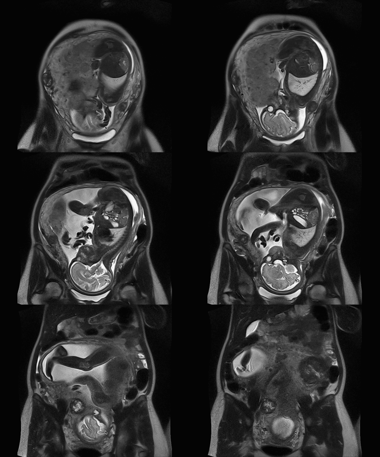
A clearer view of your pregnancy
An in-depth, safe fetal MRI that complements your routine prenatal care, with all results in one secure app
One place
A secure app for your pregnancy scans, labs and notes

In-depth
Fetal MRI
A cutting-edge
way to see
your pregnancy
Health record
Lifetime
Start your baby’s health story with a complete record of your pregnancy
Care
Effortless
Your entire pregnancy, effortlessly managed in one simple app
Join the waitlist
Be among
the first
families

A new way to see
your pregnancy
How it works
Three simple steps
A lifetime of data
The journey simply explained and it starts with a single scan
01

A safe MRI scan
Non-invasive, no ionizing radiation and no contrast agent
02

A detailed report
For you, and shared with your OB-GYN
03

All in one place
Your prenatal results on one secure platform
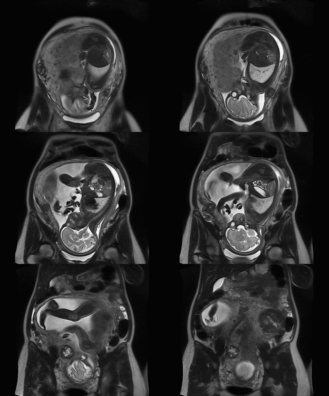
A new dimension
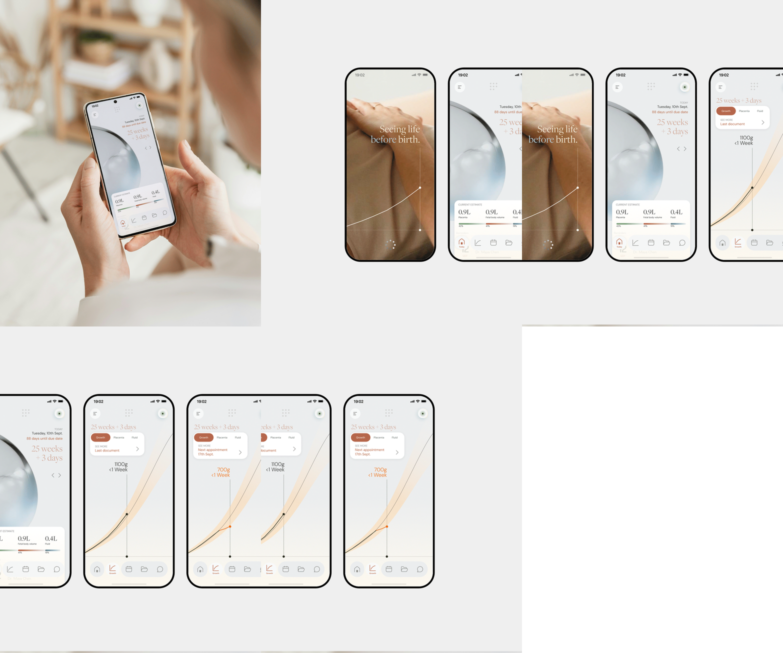
For
Two perspectives, one shared journey
One source of data, two different stories. One of knowledge. One of discovery.
For parents
Be first to try advanced pregnancy follow-up
- Book, scan, get your report, shared with your doctor.
- A short fetal MRI for a detailed view of your baby’s development, beyond routine check-ups.
- Join the waitlist for priority access and an early-bird rate.
For OB-GYNs
Join our founding clinician network.
- Your patients choose the scan. You get the full data, with no prescription and no change to your workflow.
- Founding clinicians get early access to sample reports and our clinical protocol.

Our team
Built by clinicians, researchers and healthcare operators.
FirstSight Labs is co-founded with fetal medicine
and pediatric radiology specialists from Necker Hospital
(France’s largest prenatal diagnosis center)
Scientific Advisory Board

Yves Ville, MD, PhD
Co-founder and Member, Scientific Board
Head of Obstetrics and Gynecology at Necker-Enfants Malades Hospital, professor at Paris Descartes University, member of the French National Academy of Medicine. Pioneer of fetal medicine and surgery alongside Michael Harrison and Kypros Nicolaides. Director of the National Reference Center for Twin-to-Twin Transfusion Syndrome.

Laurent Salomon, MD, PhD
Co-founder and Member, Scientific Board
Senior consultant in Obstetrics and Fetal Medicine at Necker-Enfants Malades Hospital, full professor at Paris Descartes University. Specialist in fetal medicine and surgery, obstetric ultrasound and MRI, twin pregnancies. PhD in Physics applied to placental imaging, MSc in Medical Statistics from the London School of Hygiene and Tropical Medicine. President of ISUOG since December 2022, member of the French National Academy of Medicine, co-author of more than 250 peer-reviewed publications, co-founder of the LUMIERE Foundation.

Guillaume Gorincour, MD, PhD
Co-founder and Member, Scientific Board
Pediatric and prenatal radiologist. Graduate of Marseille Medical School, professor since 2013, fellowship in Montreal. PhD in Medical Ethics, pioneer of Post-Mortem Imaging in France. More than 180 scientific publications. Practices in private setting at IMAGE2 in Marseille, affiliated with Université Paris Cité for fetal imaging research, and scientific director of IMADIS teleradiology.

David Grevent, MD
Co-founder and Member, Scientific Board
Pediatric radiologist at Necker-Enfants Malades Hospital, AP-HP. Co-lead of the LUMIERE platform, dedicated to research in fetal and placental imaging, inaugurated at Necker in 2019.

Laurence Bussières, MD
Co-founder and Member, Scientific Board
Biologist at Necker-Enfants Malades Hospital, AP-HP, founding member of the LUMIERE Foundation.

Leadership

Amine Bouachba – MD
CEO & Co-founder

Frequently Asked Questions
How does it work?
Sign up in our app, choose your membership, then book your scan with us. The MRI takes about 30 minutes, and you receive your results in under a week, often sooner. The healthcare professionals who follow your pregnancy (your OB/GYN, midwife or family doctor) receive them too.
Do I need a doctor's order?
No, you don’t need to ask your doctor for a prescription, our medical team takes care of it. We will ask for the name of your OB/GYN or midwife so we can inform them and include them in your follow-up.
Is MRI safe for my baby?
Yes, MRI uses a magnetic field and radio waves, not X-rays, so there is no radiation. One study covered 1.4 million births and followed children up to age 4. The 1,737 babies exposed to MRI early in pregnancy showed no increase in birth defects, cancer, or vision or hearing loss. Another study followed 131 exposed children and 771 unexposed children up to age 6, and their development was the same. The major obstetric guidelines agree that MRI without contrast dye has no known harmful effect at any stage of pregnancy, and we never inject contrast dye (Ray, JAMA, 2016; Zvi, AJNR, 2020; ACOG, Obstet Gynecol, 2017; Prayer, Ultrasound Obstet Gynecol, 2023).
Can the noise hurt my baby's hearing?
No, the machine is loud for you, so you get earplugs or headphones, but your body and the amniotic fluid soften the sound for your baby. In a study of 751 newborns scanned in the womb, none had hearing loss, compared with 0.34% of 10,042 babies who were never scanned. The results were the same with the most powerful 3 Tesla scanners, and hearing was still normal when children were checked again at around age 2 (Strizek, Radiology, 2015; Chartier, AJR, 2019; Bouyssi-Kobar, Pediatr Radiol, 2015).
When in pregnancy can I have the exam?
You can have it from about 18 weeks until the end of pregnancy, and our growth charts for the baby, the placenta and the amniotic fluid cover 16 to 36 weeks (Prayer, Ultrasound Obstet Gynecol, 2023; Bouachba, Ultrasound Obstet Gynecol, 2025).
Do I need to prepare?
Usually not, just wear comfortable clothes without metal, remove your jewelry, and empty your bladder before the scan. Tell us if you have an implant, a pacemaker or any metal fragment in your body.
I'm claustrophobic. Can I still have it?
Yes, just tell us when you book. The team talks with you throughout the scan, you hold a call button the whole time, and you can stop whenever you need to.
What if my baby moves a lot?
That’s perfectly normal, each image is taken in less than a second, which freezes the movement. If an image is blurred, we simply take it again.
Can my partner come with me?
Yes, your partner is welcome to come with you, but usually cannot enter the MRI room itself. You are never alone, since you can talk with the team at any moment through an intercom and healthcare professionals watch over you for the whole scan.
What does this exam tell us that ultrasound doesn't?
Think of measuring the water in a glass. Ultrasound is like guessing the amount from a photo, while MRI is like pouring it into a measuring cup. Ultrasound estimates your baby’s weight from a few flat measurements and is off by 6 to 11% on average, while MRI measures the whole volume in 3D and is off by only 3 to 4%. Even when every woman is scanned late in pregnancy, ultrasound still misses about 4 in 10 babies who are too small and 6 in 10 who are too large. It also raises false alarms: when it flags a baby as very large, it is wrong about 2 times out of 3. In a study of 2,378 pregnancies, MRI at 36 weeks detected 80% of very large babies, compared with 59% for ultrasound. Our own research built MRI growth charts for the baby, the placenta and the amniotic fluid, which ultrasound can only estimate indirectly (Kadji, Am J Obstet Gynecol, 2019; Kadji, Am J Obstet Gynecol, 2022; Sovio, Lancet, 2015; Sovio, Ultrasound Obstet Gynecol, 2018; Bouachba, Ultrasound Obstet Gynecol, 2025; Bouachba, Ultrasound Obstet Gynecol, 2026; Bouachba, Acta Obstet Gynecol Scand, 2026).
Does it replace my ultrasounds?
No, it adds to your usual prenatal care, so keep all the visits and ultrasounds your doctor has planned. MRI is highly precise on its own, and combining it with your ultrasound measurements, lab results and medical history gives an even more complete picture of your baby’s growth. You’ll find all of this together on your app dashboard, a step toward truly personalized prenatal care.
What if a result is outside the usual range?
A number outside the range is not a diagnosis, and many healthy babies are simply smaller or larger than average. If the radiologist suspects a problem, your OB/GYN is notified right away so you get the right care. Your doctor will explain what the result means for you and decide on any next steps. Our analysis supports your doctor’s judgment and never replaces it.
Can we see the images?
Yes, anytime. Our app brings your MRI, ultrasounds and lab results together in one place, shows your baby as a 3D avatar, and lets you follow your baby’s growth throughout your pregnancy.
Is it possible with twins?
Yes, absolutely, MRI works for twin pregnancies too, and our analysis covers them.
How much does it cost? Is it covered by insurance?
You’ll find our membership options on the Pricing page. Most insurance plans do not cover the exam yet, but you can usually pay with your HSA or FSA (Health Savings Account or Flexible Spending Account), because diagnostic medical care is an eligible expense. Check with your plan administrator to confirm (IRS, Publication 502, 2025; IRS, Publication 969, 2025).
How is my data protected?
Your health information is protected under HIPAA, the U.S. federal privacy law for medical data. We never sell your data, and your images are used for research only if you give your explicit consent (HHS, Summary of the HIPAA Privacy Rule).
This page provides general information and does not replace advice from your doctor or midwife.
Your baby’s story starts now
Get early access
Join the waitlist to be notified when a center opens near you.
By clicking Join you’re confirming that you agree with our Terms and Conditions.